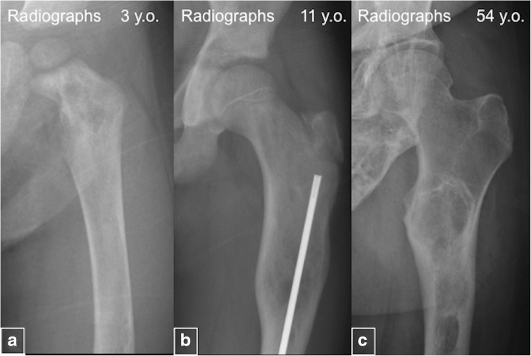
Fig. 12.
“Evolution” of the fibrous dysplasia (FD) lesions. a Radiograph of a 3-year-old demonstrates a typical heterogeneous-appearing FD lesion in the femur. b Radiograph from an 11-year-old demonstrates homogeneous and radiolucent FD lesion. c Image from a 54-year-old patient shows sclerotic FD lesions

