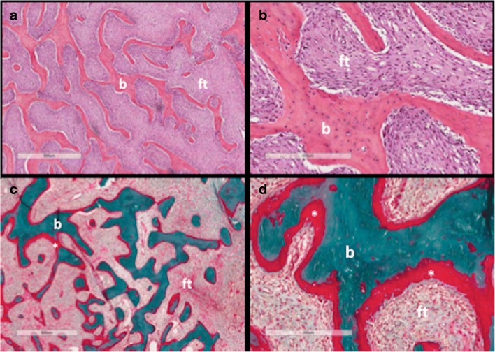
Fig. 3.
Histopathological features of fibrous dysplasia (FD). FD lesions are composed of fibrous tissue interspersed between bone trabeculae. The amount of bone within lesions is quite variable. Trabeculae are dysplastic, non-stress oriented, and appear disorganised. Haematoxylin-eosin stained sections in low (a) and high power (b) show irregular, discontinuous trabeculae (b) within a fibrous stroma (ft), demonstrating the typical “alphabet soup” pattern. Goldner’s trichrome stained sections in low (c) and high power (d) reveal osteomalacic changes including excess osteoid (asterisks) and severe undermineralisation of the dysplastic bone (reprinted from Boyce [34])

