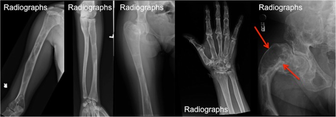
Fig. 5.
The radiographic appearance of fibrous dysplasia (FD) and the rind sign. a–e Frontal radiographs demonstrate classic FD lesions in appendicular skeleton. A classic lucent lesion surrounded by a layer of sclerotic reactive bone (so-called the rind sign). The rind sign is most commonly seen in the proximal femur (red arrow)

