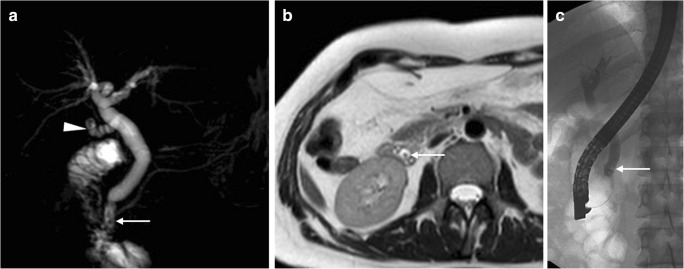
Fig. 15.
Residual lithiasis in the common bile duct (CBD) following recent (10 days) laparoscopic cholecystectomy performed at another hospital. MRCP (a) and axial T2-weighted (b) MR images showed unremarkable cystic duct remnant (arrowhead) and mildly dilated CBD with small dependent filling defects (thin arrows). Residual lithiasis was confirmed at ERCP (thin arrow in c) and treated by sphincterotomy and extraction using a Dormia basket

