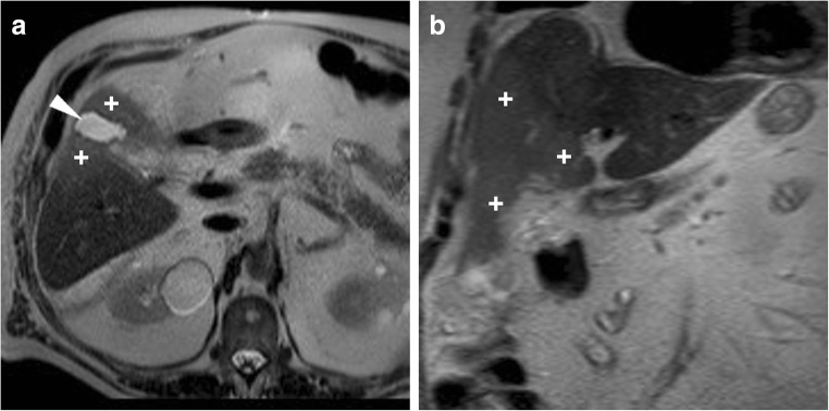
Fig. 8.
Expected MR appearance 24 h after uncomplicated laparoscopic cholecystectomy: axial (a) and coronal (b) T2-weighted images showing small-sized fluid collection in the gallbladder fossa, surrounded by increased signal intensity of the liver parenchyma (+), reflecting transient postoperative hepatic oedema

