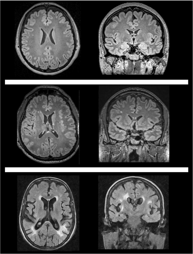
Fig. 2.
White matter involvement in Fabry disease could range from small, scattered and punctuate T2-weighted hyperintense foci (upper row, 46-year-old woman, or middle row, 52-year-old man) to bilateral diffuse, patchy and partly confluent white matter hyperintensities (lower row, 40-year-old woman). Although being the most common neuroimaging finding in Fabry disease, white matter hyperintensities appearance and distribution are not specific in this condition

