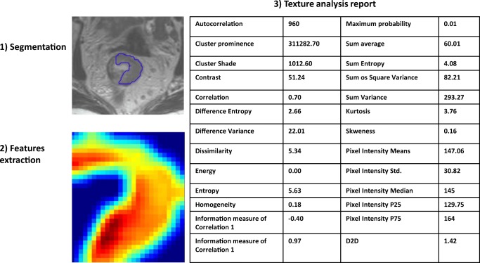
Fig. 1.
Example of texture analysis in MRI of rectal cancer performed with QUIBIM software (QUIBIM S.L., Valencia, Spain). The region of interest for the texture is defined by manual segmentation (1). The texture model is extracted by the software through a grey-level co-occurrence matrix analysis (2) that enables the extraction of a set of features that are shown in a structured report (3). The same region of interest can be used to extract other features based on intensity histogram, shape, and so on.

