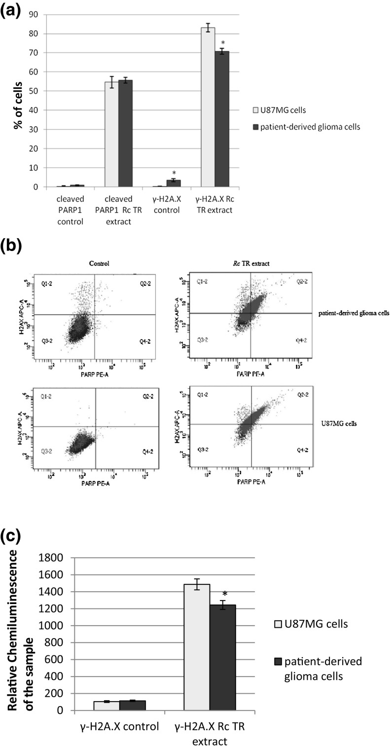
Fig. 3.
Rc TR extract increased the numbers of cleaved PARP1- and γ-H2A.X-positive cells. a The graph presents the percentage of cleaved PARP1- and γ-H2A.X-positive patient-derived glioma cells and U87MG cells measured by flow cytometric analysis after 24 h treatment with Rc TR extract. b Representative flow cytograms. Control sample cells of both patient-derived glioma cells and U87MG cell line are located predominantly in bottom left quarter of the graph (cleaved PARP-negative, γ-H2A.X-negative). After treatment with Rc TR extract both samples appear at PAPR-positive and γ-H2A.X-positive areas of the graph indicating growth of dead cell population and ongoing DNA-repair processes. c The graph presents the level of γ-H2A.X after 24 h treatment of patient-derived glioma cells and U87MG cells with Rc TR extract, measured by Elisa test. Results represent mean ± SD from 3 independent experiments. *p < 0.05 patient-derived glioma cells versus U87MG cells

