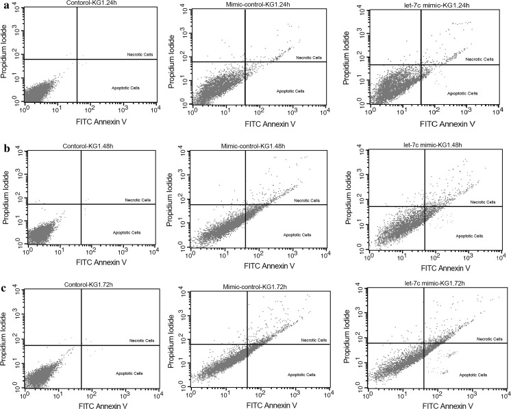
Fig. 4.
Evaluation of apoptosis and necrosis in the untreated cells, mimic control and hsa-let-7c mimic groups by Annexin-V and propidium iodide (PI) staining performed 24, 48 and 72 h after transfection (a–c). Flow cytometry analysis was performed using 488-nm excitation and a 515-nm band-pass filter for fluorescein detection and a 600-nm filter for PI detection

