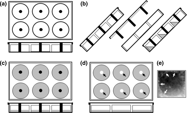
Fig. 3.
Schematic representation of modified-6-well culture plate application in the s-ARU method for migration assay using pipette tips as a barrier. a Upper and lateral view of the modified-6-well culture plate. b Lateral view of the tilted modified-6-well culture plate for application of the medium-containing cells. c Upper and lateral view of modified-6-well culture plate in horizontal position, allowing cells to attach and to reach confluence. At this moment, cover plate needs to be fixed with the help of sterile paper tape to restrict the free movement of the cover plate and preventing disturbance of the free-cell borderline. d Upper and lateral view of culture plate after cells have reached the confluence. A regular cover plate replaces the modified cover plate. The arrows point to the cell-free gap formed at the center of the wells. e Representative view of the cell-free gap formed by LNCaP cells after zone formation under normal condition. Arrows point to the borderline of the cell-free gap

