Fig. 5.
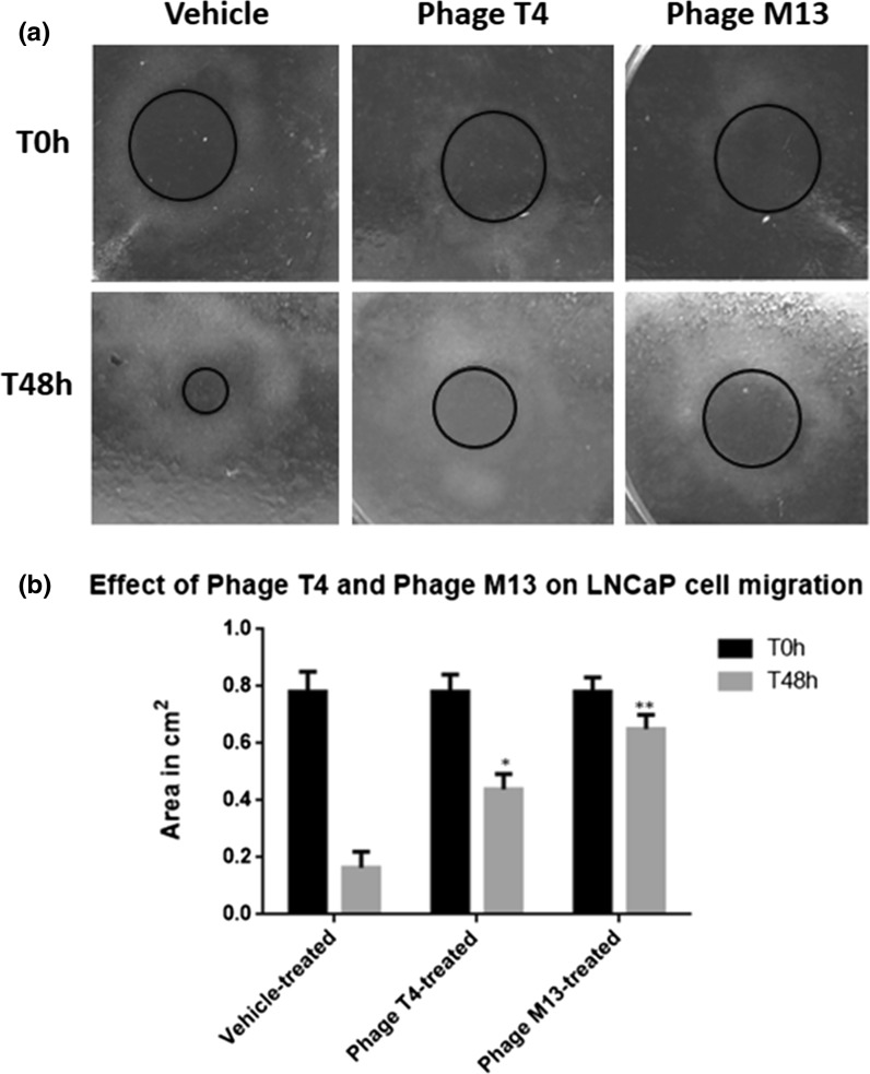
Representative image of LNCaP cells cultured using pipette tips as a barrier (s-ARU method). a After the incubation of LNCaP cells with pipette tip barrier for 24 h, the cell-free gap could be easily identified (red circle), for all treatment, and it represents time zero hour (T0 h) or the beginning of the experiment. LNCaP cells were exposed for 48 h (T48 h) to bacteriophages T4 and M13 at 1 × 10⁷ pfu/ml concentration. b Results of the measurements of the cell-free area (red circle) after 48 h (T48 h) of LNCaP cell growth and treated with bacteriophages T4 and M13. Note that both phages reduced the rate of LNCaP cell migration, in which phage M13 (** means p < 0.001) was more effective as compared with the phage T4 (* means p < 0.01). The values are presented as the mean ± standard deviation. (Color figure online)

