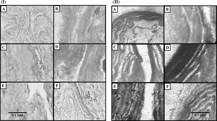
Fig. 4.
I Cell adhesion and growth visualization by H&E staining. a Control scaffold, b PRP coated scaffold. c Scaffold treated with osteogenic medium. d PRP coated scaffold treated with osteogenic medium. e Scaffold treated with PRP 5%. f PRP coated scaffold treated with PRP 5 %. ×40 magnification. II Alizarin red staining confirms the osteogenic differentiation of all groups. a Control scaffold, b PRP coated scaffold. c Scaffold treated with osteogenic medium. d PRP coated scaffold treated with osteogenic medium. e Scaffold treated with PRP 5%, f PRP coated scaffold treated with PRP 5%

