Figure 1.
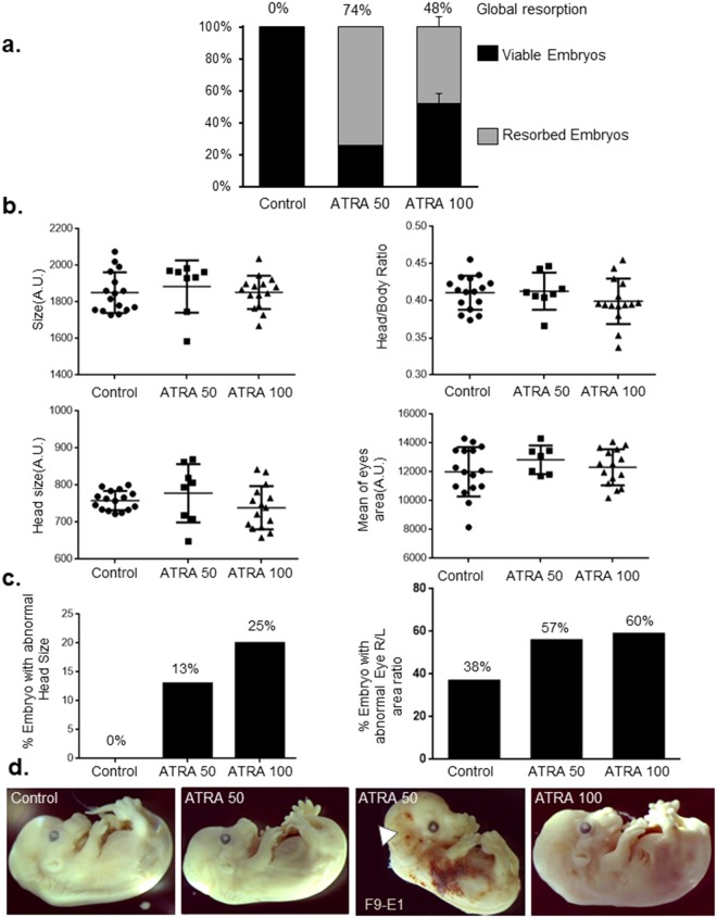
(a) Analysis of the embryonic resorption after E9.5 intraperitoneal mice injection of ATRA at 50 mg/kg (one experiment), at 100 mg/kg and control condition (DMSO) (2 independent experiments). Viable embryos are represented in black bars whereas resorbed embryos are in gray bars. (b) Head, eye and body size measurements of treated and control embryos at E14.5. No statistical differences were observed concerning global parameters such as embryo size, head size, ratio of size of head and body, area of eyes. (c) Percentage of embryos presenting abnormal Head size (left) and eyes area ratio (right). The 90% confidence interval on the mean was computed for each group. Each embryo with a measurement outside the 90% confidence interval was considered with abnormal item. The percentage of embryos with an abnormal head size or abnormal eyes area ratio is calculated (right eye area divided by the left eye area). (d) Representative embryos per condition at E14.5. Treated embryos were not, at the first look, especially malformed compared to controls even if hemorrhages have been observed most often in treated embryos. Notably, a bilateral anotia (white arrow) is observed in F9-E1 embryo (ATRA-50). This embryo was excluded of quantitative analysis due to absence of several craniofacial structures (ear, Meckel’s cartilage). n = 16 embryos from 2 females were analyzed for the control group, n = 8 embryos from one female for ATRA 50 and n = 15 embryos from 2 females for ATRA 100.

