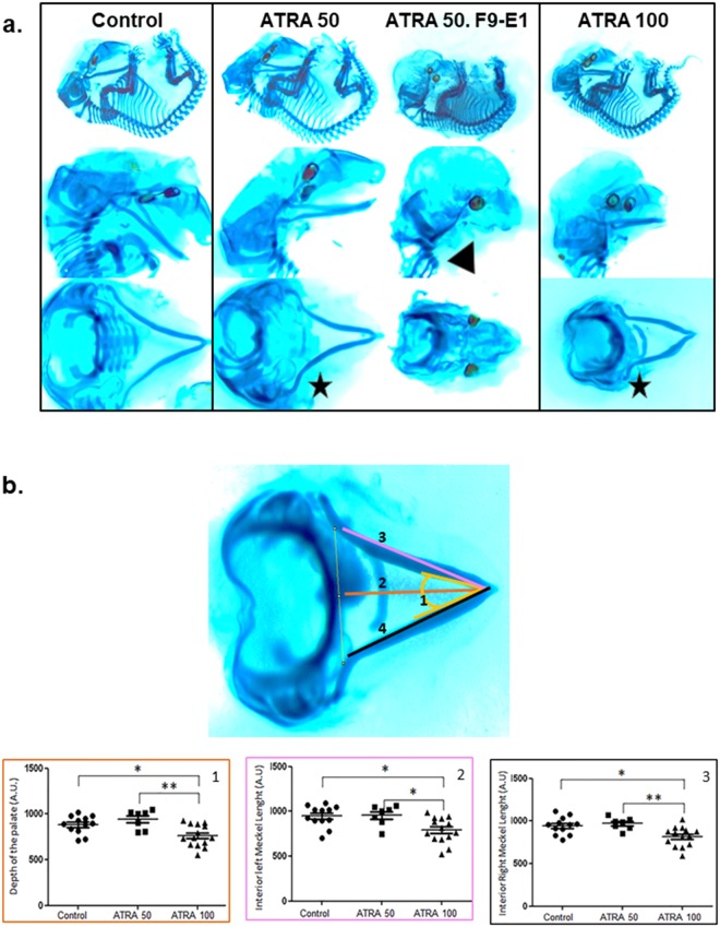
Figure 2.
(a) Craniofacial cartilages of embryo staining with Alcian Blue-Alizarin Red coloration. Differences in size and/or angle of the Meckel’s cartilage were observed (black stars) after ATRA treatment (50 and 100 mg/Kg). The F9-E1 embryo (ATRA 50) presented a total aplasia of this cartilage (black arrow). This embryo was excluded of quantitative analysis due to absence of several craniofacial structures. (b) Quantitative analysis of several craniofacial cartilage parameters. The localization of these 4 different parameters was detailed in Alcian Blue-Alizarin Red staining (up-left panel). ANOVA revealed significant group effect for the depth of the palate (1), F(2) = 6.136 with p = 0.0058; the left length (2) and right (3) length of the Meckel’s cartilage and also, F(3) = 6.453with p = 0.0047; (F(4) = 7.264 with p = 0.0027; Post-hoc analysis by Tukey *p < 0.05, **p < 0.01, ***p < 0.001 were then performed. For Control, n = 16 embryos from 2 females were analyzed, n = 8 embryos from one female for ATRA 50 and n = 15 embryos from 2 females for ATRA-100). For ATRA 100 treatment, the studied parameters are significantly different from control, and then may reveal a real macroscopic effect.

