Fig. 2.
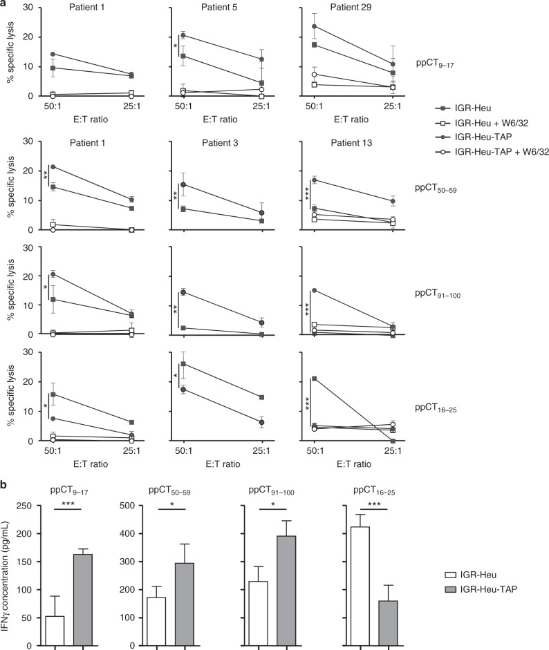
Specificity of ppCT-peptide-stimulated CD8+ T cells. a NSCLC patient PBMCs were stimulated in vitro with the indicated peptides, and then CD8+ T cells were isolated and their cytotoxic activity was tested. The IGR-Heu and IGR-Heu-TAP (IGR-Heu transfected with TAP1 and TAP2) tumour cell lines, generated from patient 1 (Heu), pre-incubated or not with neutralizing anti-MHC-I mAb W6/32, were used as targets. Cytotoxicity was determined by a conventional 4-h 51Cr release assay at the indicated E:T ratios. Values correspond to means (±SD) of percentages of lysis from triplicates. Data shown represent experiments from three patients' PBMCs. b Cytokine release of patient 1’s T cell clones and T cell cloids stimulated with the autologous IGR-Heu and IGR-Heu-TAP cell lines. IFN-γ release was measured using ELISA. Data shown are means (±SD) of three T cell clones and three T cell cloids from three independent experiments. *p < 0.05; ***p < 0.001 (two-tailed Student’s unpaired t test). E:T effector:target

