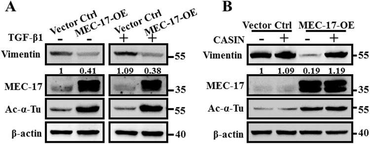
Figure 7.
Pharmacological inhibition of cdc42 activity rescued MEC-17-induced suppression of EMT. (A) The representative Western blots show the protein levels of vimentin, MEC-17, and acetyl-α-tubulin in vector control and MEC-17-overexpressed A549 cells after treatment with TGFβ1 (20 ng/mL) for 48 h. Beta-actin served as the internal control. The relative vimentin intensities normalized to vector control group were shown. Uncropped blots are displayed in the supplementary information (Supplementary Fig. S5). (B) The representative Western blots show the protein levels of vimentin, MEC-17, and acetyl-α-tubulin in vector control or MEC-17-overexpressed A549 cells after treatment with the cdc42 inhibitor CASIN (5 μM) for 48 h. Beta-actin served as the internal control. The relative vimentin intensities normalized to vector control group were shown. Uncropped blots are displayed in the supplementary information (Supplementary Fig. S5).

