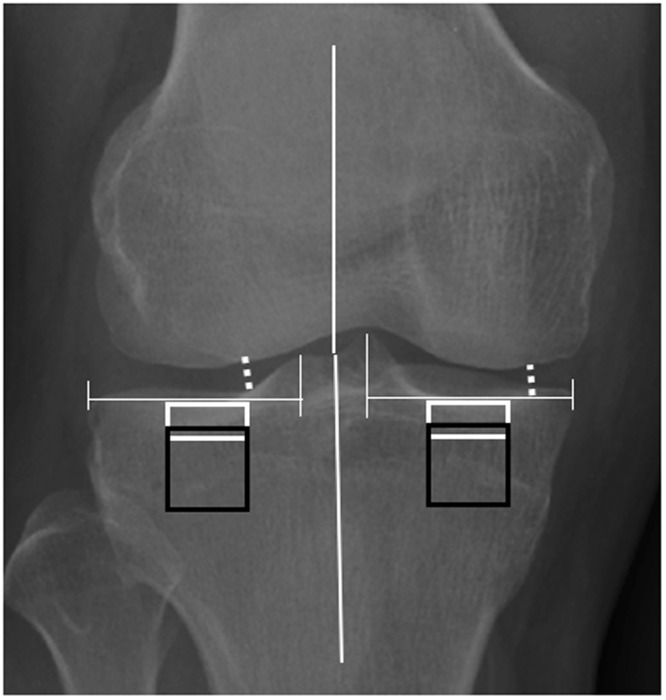
Figure 5.

Schematic figure of placement ROIs. Two subchondral bone ROIs (93 × 40 pixels, white rectangles) were placed under the cartilage-bone interface in the middle part of medial and lateral condyles of the tibia. Two trabecular bone ROIs (93 × 93 pixels, black rectangles) were placed below and parallel to the subchondral bone ROIs of the tibia. Minimum JSWs (white dotted line) were measured from the narrowest point of the joint from both the medial and the lateral sides. Anatomical angles were medially measured from the intersection of a line from the center of the head of femur to the center of the tibial spines, and a second line from the center of the tibia to the center of the tibial spines.
