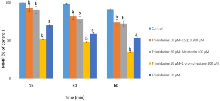
Figure 3.
Hepatocytes (106 cells/mL) were incubated in Krebs–Henseleit buffer, pH 7.4, at 37 °C for 60 min following the addition of thioridazine (50µM). Mitochondrial membrane potential was determined as the percentage of mitochondrial rhodamin reuptake between control and treated cells (9). GSH depleted hepatocytes (with 1-bromoheptane) were prepared as described by Khan et al. (21). Values are expressed as mean ± SEM of three separate experiments
a Significant difference in comparison with control hepatocytes (p < 0.05).
b Significant difference in comparison with thioridazine-treated hepatocytes (p < 0.05).

