Figure 1.
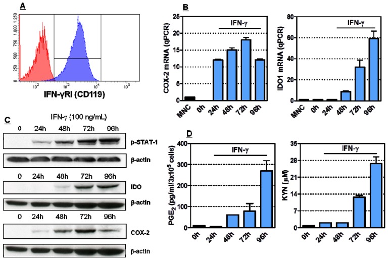
Induction of COX-2 and IDO1 in HL-60 leukaemia cells. Panel A: The expression of IFN-γ receptor I (CD119) was investigated by flow cytometry. One representative experiment out of 4 with similar results is shown. Panel B: Quantitative RT-PCR was conducted to measure COX-2 and IDO1 mRNA levels in IFN-γ challenged HL-60 leukaemia cells. Graphs summarize 5 independent experiments. Data are expressed in terms of mean ± SEM. Panel C: HL-60 cells were activated with 100 ng/mL IFN-γ for up to 96 h, followed by Western blot runs to detect phosphorylated STAT1, COX-2 and IDO1 proteins. Panel D: Measurement of PGE2 and kynurenines in supernatants of HL-60 leukaemia cells stimulated with 100 ng/mL IFN-γ. Bars reflect the mean and SEM recorded in 3 independent experiments.

