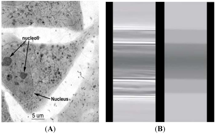
Figure 1.
(A) Cryo X-ray microscopy of 3T3 cells [31]. (B) Direct comparison between the phase-contrast radiological image based on coherence of an optic fiber (on the left) and the corresponding absorption-contrast image (right) [34]. Adapted by permission from John Wiley and Sons and IOP Publishing.

