
Figure 4.
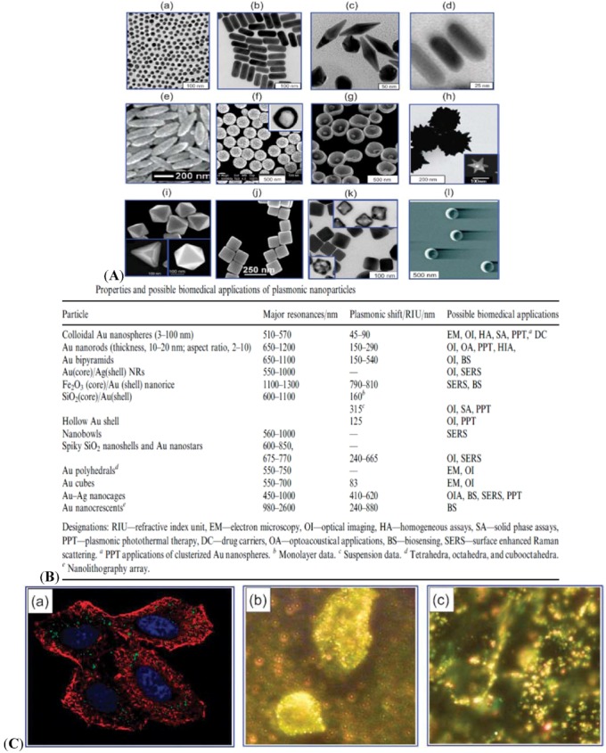
(A) Various types of plasmon-resonant nanoparticles: 16 nm nanospheres (a) Au nanorods (b) Au bipyramids (c) Au nanorods surrounded by silver nanoshells (d) nanorice (Au-coated Fe2O3 nanorods) (e) SiO2/Au nanoshells (f) (the inset shows a hollow nanoshell); nanobowls with bottom cores (g) spiky SiO2/Au nanoshells (h) (the inset shows a Au nanostar); Au tetrahedra, octahedra, and cubooctahedra (i) Au nanocubes (j) Ag nanocubes and Au-Ag nanocages (obtained from those in the insets) (k) as well as Au nanocrescents (l) [9]. (B) Properties and possible biomedical applications of plasmonic nanoparticles. (C) Confocal image of HeLa cells in the presence of AuNPs [77]. (a) Blue indicates the nuclei stained with Hoechst 33258. Red indicates the actin cytoskeleton labeled with Alexa Fluor 488 phalloidin. Green indicates unlabeled AuNPs. The image is taken by two-photon microscopy. Dark-field microscopy of cancerous (b) and healthy (c) cells using AuNPs conjugated with antibodies for epidermal growth factor [78]. Adapted from the data of the cited papers by permission from The Royal Society of Chemistry, and The American Chemical Society.