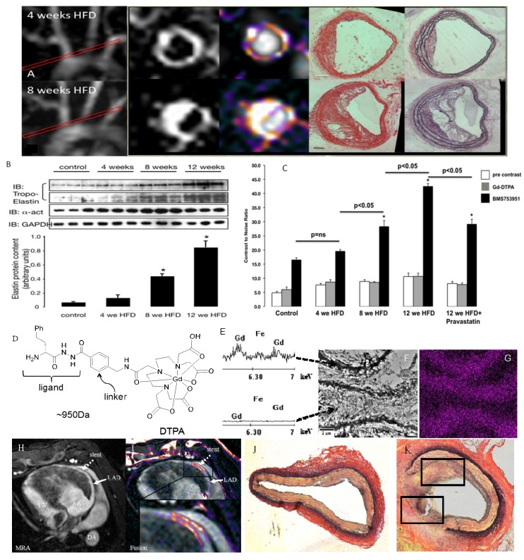
Figure 9.
(A) MRI of extracellular matrix remodeling in an apoE−/− mouse model of accelerated atherosclerosis at 4 and 8 weeks after commencement of HFD using an elastin specific MR contrast agent (D), ESMA (Lantheus Medical Systems, North Billerica, MA, USA). (B,C) Contrast-to-noise values after ESMA injection increased with the duration of the HFD, which was paralleled by an increase in tropoelastin by western blotting. Electon microscopy of elastin fibers (F) and X-ray spectroscopy of gadolinium (Gd) (E,G) showed preferential uptake of ESMA along the elastic fibers with little to no uptake in-between elastin fibers (F,G). (F) ESMA MRI of mechanical coronary wall injury after MR lucent stent placement (H) showed focal signal enhancement in the stented area (I), which was in agreement with extracellular matrix remodeling on histology (J,K) (adapted from Makowski [80] and von Bary [81]).

