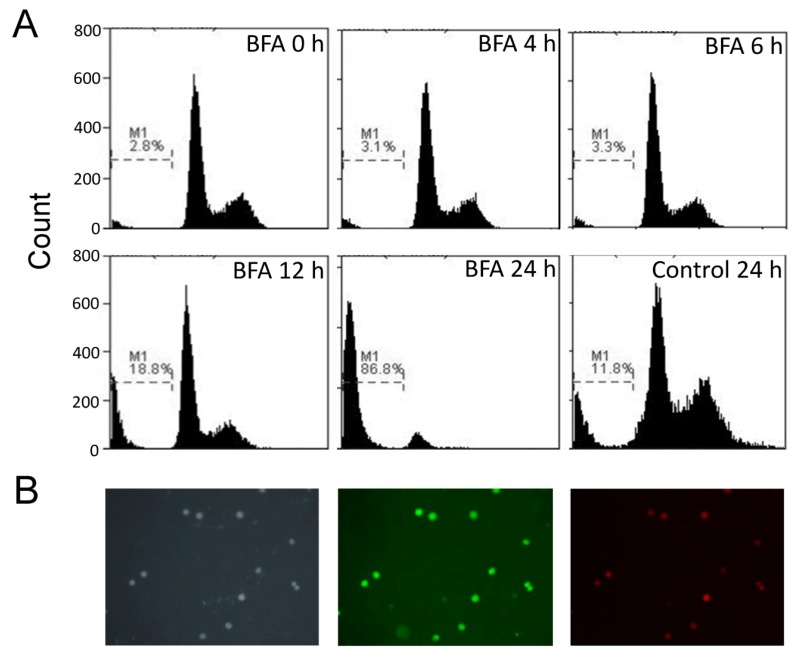
Figure 4.
The BFA induced apoptosis of Colo 205 cells in suspension cultures. (A) Colo 205 suspension cells were treated with 0.1 μg/mL BFA for 0, 4, 6, 12 and 24 h. The cells were then harvested, fixed and stained with propidium iodide for flow cytometry assay. Sub-G1 region (M1) represented apoptotic cells. (B) Suspension Colo 205 cells treated with 0.1 μg/mL BFA for 12 h were stained with DAPI (2-(4-Amidinophenyl)-6-indolecarbamidine dihydrochloride, left panel), Annexin V-FITC (middle panel) and propidium iodide (right panel), and photographed under a fluorescence microscope at 100× magnification. Representative results from two independent experiments were shown.

