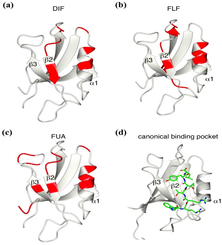
Figure 3.
Identification of the interface between the mZO-1 PDZ1 domain and a compound. Mapping of residue signal changes upon mixing with DIF (a), FLF (b), and FUA (c) onto the ribbon model of the mouse ZO-1 PDZ1 domain (PDB:2RRM). (d) The ribbon model represents the canonical binding pocket between the PDZ domain and peptide (PDB:2H2B).

