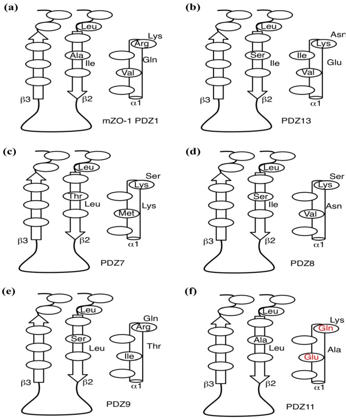
Figure 4.
Comparison of the interfacial residues. Schematic of the interfacial residues of mZO-1 PDZ1 (a), PDZ13 (b), PDZ7 (c), PDZ8 (d), PDZ9 (e), and PDZ11 (f) are depicted. The canonical binding pocket lies between a β-sheet composed of β2 and β3 strands and α1 helix, represented as block arrows (β2 and β3 strands) and a cylinder, respectively. Each ellipse represents the location of each residue. The atypical residues of PDZ11 are in red.

