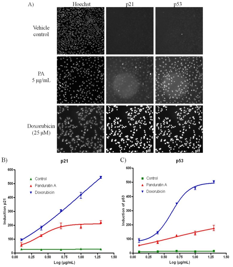
Figure 5.
PA induced oxidative stress on A549 cells. (A) Fluorescence images acquired on the ArrayScan HCS Reader showing induction of p53 and p21 in the nucleus of A549 cells after 20 h incubation. Doxorubicin was included as positive control. While the untreated cells have minimal p53 or p21 signal, doxorubicin treated cells show bright p53 and p21 labeling in the cell nuclei; (B) PA induced p21 was evidenced with increased of the fluorescence signal; (C) PA induced p53 was evidenced with increased of the fluorescence signal.

