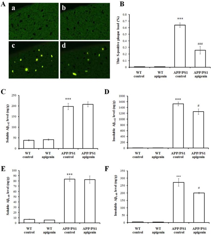
Figure 2.
Apigenin treatment relieves Aβ burden in APP/PS1 mice. (A) Representative images of Thio S positive compact plaques staining in cortex of wild-type mice and APP/PS1 mice (400 ×). a, WT vehicle control; b, apigenin-treated WT; c, APP/PS1 vehicle control; d, apigenin-treated APP/PS1. (B) Quantitative analysis of Thio S positive compact plaques staining. *** p < 0.001 vs. vehicle-treated WT mice, ### p < 0.001 vs. APP/PS1 control mice. Data are presented as mean ± S.E.M., n = 4. (C–F) ELISA analysis of soluble or insoluble Aβ1–42 (C,D), 1–40 (E,F) levels in extracts of cerebral homogenates of WT and APP/PS1 mice. *** p < 0.001 vs. WT control mice, # p < 0.05 vs. APP/PS1 control mice. All data are presented as mean ± S.E.M., n = 4.

