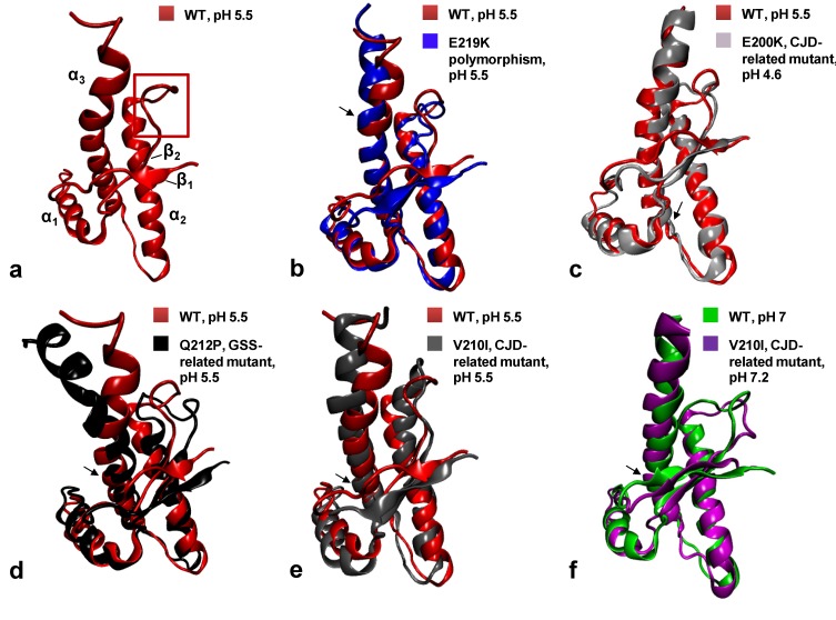
Figure 2.
Cartoon representations and backbone heavy atom overlays of the different WT and mutants HuPrP folded domains (residues 126–231): WT at pH 5.5 in red (a, PDB code 2LSB), E219K in blue (b, PDB code 2LFT), E200K in light gray (c, PDB code 1FO7), Q212P in black (d, PDB code 2KUN), V210I at pH 5.5 in gray (e, PDB code 2LEJ) and (f) V210I at pH 7.2 in magenta (PDB code 2LV1) superimposed with the WT at pH 7 in green (PDB code 1HJN). The red frame on the WT (a) highlights the β2-α2 loop region. Black arrows point at the location of each mutation.

