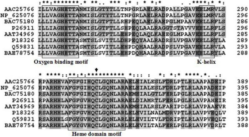
Figure 1.
Cytochrome P450 alignments of the amino acid sequence with other bacterial strains. P26911; CYP from Streptomyces griseus, BAC75180; CYP from Streptomyces avermitilis MA4680, AAC25766; CYP from Streptomyces lividans, NP_625076; CYP from Streptomyces coelicolor A3(2), AAT34969; CYP from Streptomyces tubercidicus, P18326; CYP from Streptomyces griseolus, BAE78754; CYP from Streptomyces sp. TM-7, Q59831; CYP from Streptomyces carbophilus. Multiple sequence alignment of conserved motifs in P450 enzymes identified. Both oxygen-binding motif and heme domain sequences as well as the EXXR motif in the K helix are shown. Amino acids conserved in the P450s listed are shown. The Cys residue that coordinates the heme is indicated by an arrow. Alignments were performed with the CLUSTAL X 1.83. (*) Identical residues in the different enzymes; (:) conserved substitutions; (.) semi-conserved substitutions.

