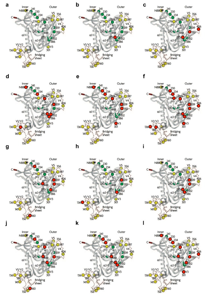
Figure 2.
Positions of mutated N-linked glycans under selective CBA pressure. (a) Ribbon diagrams show the 24 N-linked glycosylation sites in recombinant (monomeric) gp120 of wild-type HIV-1 IIIB according to Leonard et al. [3] and Kwong et al. [11]. The green dots indicate the high-mannose type glycans and the yellow dots the hybrid/“potential” complex types. Recent findings by Doores et al. [8] and Bonomelli et al. [9] demonstrated that the glycan composition of recombinant monomeric gp120 differs greatly from that of trimeric gp120 present on infectious viral particles, of which the latter were used in the described resistance studies. Based on these data, the specific glycosylation assignments presented here are an overestimate of complex glycan content. Positions of the glycans deleted on gp120 under increasing concentrations of the following CBAs: (b) 2G12 mAb (NL4.3), (c) 2G12 mAb (IIIB), (d) HHA (IIIB), (e) GNA (IIIB), (f) AH (IIIB), (g) CV-N (IIIB), (h) CV-N (NL4.3), (i) MVN (NL4.3), (j) BanLec (IIIB), (k) GRFT (IIIB) and (l) UDA (IIIB) are marked with red dots. Figure 2a, reproduced with permission, from reference [12].

