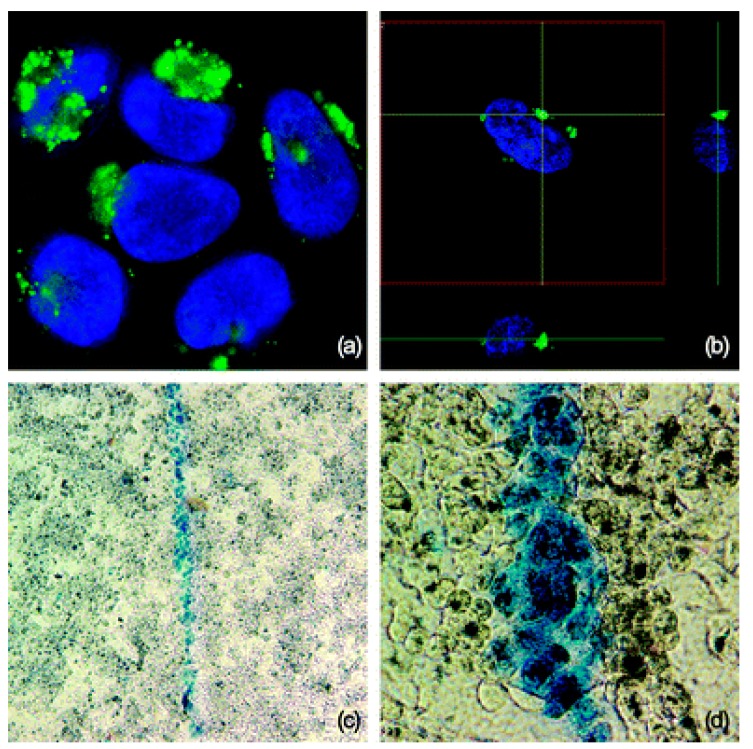
Figure 2.
(a) Representative confocal microscope picture of lung A549 cells labeled with the FITC doped GSS nanoparticles showing the presence of nanoparticles (green) near the nucleus (blue—stained with Hoechst). (b) A z-position cross section showing the localization of GSS nanoparticles adjacent to the nuclear boundary. (c) Trypan blue stained dead cells as ablated selectively along the path of the NIR laser and unharmed surrounding cells. (d) Higher magnification of trypan blue stained dead cells (reproduced from [38] with permission from The Royal Society of Chemistry).

