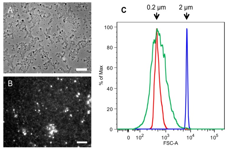
Figure 4.
Comparison of SSCs and 2 k chitin particles from C. albicans yeast cells.Microscopic analysis of (A) 2 k chitin viewed by phase contrast microscopy; and (B) SSCs viewed by fluorescence microscopy after CFW staining; (C) Flow Cytometry analysis of SSCs. Red and blue indicate the profiles of 0.2 and 2 µm standard beads, respectively; green indicate the profile of SSCs. Bars, 10 µm.

