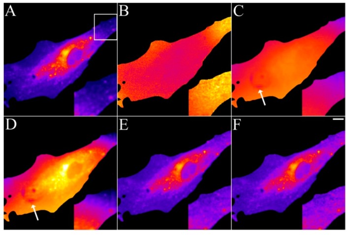
Figure 3.
Removal of background and illumination shading in DHE-labeled fibroblasts. Human fibroblasts were labelled with DHE/MCD for 3 min, followed by a chase in buffer medium for 1 h at 37 °C. Bleach stacks were acquired on a UV-sensitive wide field microscope and fitted on a pixel-by-pixel basis to a mono-exponential decay function in PixBleach. The fit provides the amplitude-map (A), the time constant map (B) and the background map. The TiEm calculated including the estimated background reveals image shading and some dark pixels stemming from the camera (arrows). The TiEm without background was either calculated from the summed pixel intensities (E) or as product of amplitude and time constant (F). Insets corresponding to the box in panel (A) illustrate the correction for illumination shading in panel (E,F). Bar, 10 μm.

