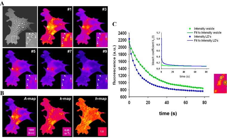
Figure 7.
Stretched exponential photobleaching of BODIPY-cholesterol in lipid droplets. HeLa cells were labelled with BChol/MCD and imaged on a wide field microscope as described in Materials and Methods. Bleach stacks were fitted on a pixel-by-pixel basis to a StrExp function implemented in PixBleach. (A) most left panel shows bright field (Bf) image of cells with zoomed box indicating LDs (arrowheads) and position of endocytic vesicles, which do not give contrast in the Bf image (arrows). Selected frames of the bleach stack shown in the next left and following frames of panel (A) reveal preferred photobleaching of BChol in LD’s compared to the rest of the cells (see especially the inset, pointing to LD’s with arrowheads and slower bleaching vesicles with arrows); (B) shows from left to right the amplitude maps (‘A-map’), the map of fitted bleach rate constants, kb, (‘k-map’), and a map of the stretching parameters (‘h-map’); (C) fit of Equation (48) to a selected LD (blue symbols, data; blue line, fit) and vesicle (green symbols, data; green line, fit); the region selected is highlighted in the corresponding colour in the zoomed inset, which is the same, as in panel A. Inset of (C) shows time evolution of the bleach rate coefficient, as defined in Equation (47) using the fitting parameters.

