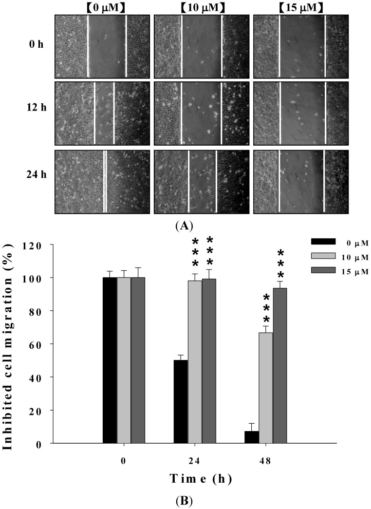
Figure 2.
Deguelin affecting the migration of U-2 OS cells examined by wound healing assay. Cells (2 × 10⁵ cells/well) were placed on the dish for 24 h before a wound was produced by scraping confluent cell layers with a pipette tip. Deguelin was added to the well at the final concentration (0, 10 and 15 μM) then incubation for 0, 12 and 24 h (A). Some representative photographs of migrating treated and untreated cells are presented. The migrated cells in the five random fields after exposure for 0, 12, 24 h were counted to quantify, and data was expressed as mean ± S.D. *** p < 0.001 significant difference between deguelin-treated groups and the untreated groups as analyzed by Student’s t test (B).

