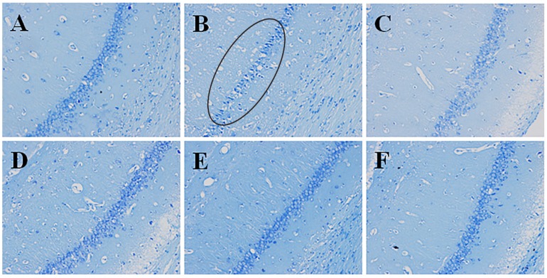
Figure 5.
Effects of pinocembrin on pathomorphological changes in the hipocampus of rats subjected to 2 h of MCAO followed by 24 h reperfusion. Representative photographs of tissue sections stained with Nissl’s staining method in the hipocampus. The pathomorphological changes in model group were marked by a cycle (B). (A) sham; (B) model (I/R); (C) I/R + pinocembrin 1 mg/kg; (D) I/R + pinocembrin 3 mg/kg; (E) I/R + pinocembrin 10 mg/kg; (F) edaravone 3.5 mg/kg. (magnification: 200×).

