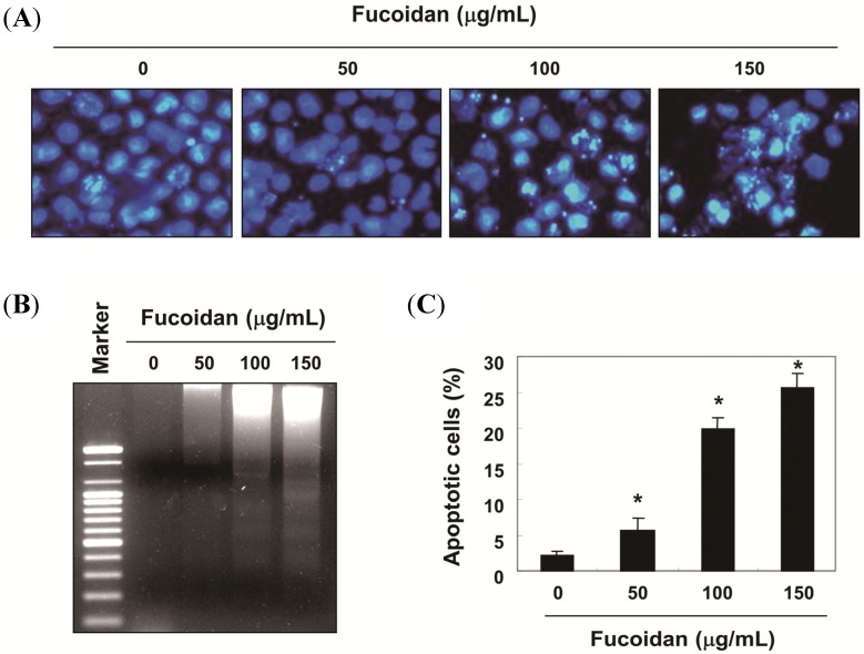
Figure 2.
Induction of apoptotic cell death of T24 cells by fucoidan treatment. (A) Cells were treated with various concentrations of fucoidan for 48 h to examine the nuclear morphological changes. The cells were fixed and stained with DAPI solution. After 10 min of incubation at room temperature, stained nuclei were then observed with a fluorescent microscope (original magnification, ×400); (B) For the DNA fragmentation analysis, genomic DNA was extracted from cells grown under the same conditions as (A) separated by 1.0% agarose gel electrophoresis, and visualized under ultraviolet (UV) light after staining with ethidium bromide (EtBr). The marker indicates a size marker of the DNA ladder; (C) To quantify the degree of apoptosis induced by fucoidan, the cells were harvested to determine the percentage of annexin V positive/propidium iodide (PI) negative (apoptotic cells). The data are expressed as the mean ± SD of three independent experiments. The significance was determined by Student’s t-test (* p < 0.05 vs. untreated control).

