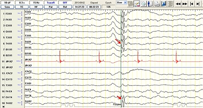
Figure 7.

Electroencephalographic recordings from a 2‐year‐old female Siberian husky (idiopathic epilepsy group) with unilateral hippocampal atrophy on the right showing polyspike activity (read arrow) from the T4 lead. The discharge localization is defined based on the highest amplitude in the reference montage (arrow) and reversed polarity in the bipolar montage recording (arrow)
