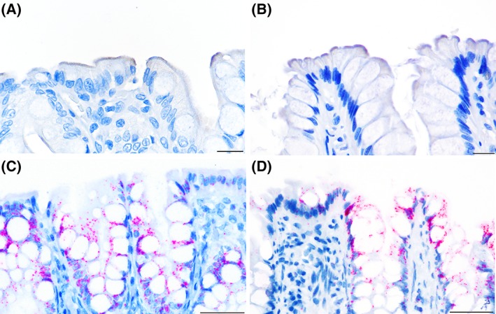
Figure 2.

Distribution of immunolabeling (in brown) for the ASBT protein (A and B) and in situ hybridization (in red) for ASBT mRNA (C and D) in the colon. In both control dogs (A) and dogs with CIE (B), the immunolabeling in the apical membrane of the superficial colonocytes was multifocal. ASBT mRNA expression was observed in both the nucleus and cytoplasm of superficial and cryptal colonocytes and was similar between control dogs (C) and dogs with CIE (D). Scale bar is equal to 15 μm, magnification of ×600 (A and B) or 50 μm, magnification of ×400 (C and D)
