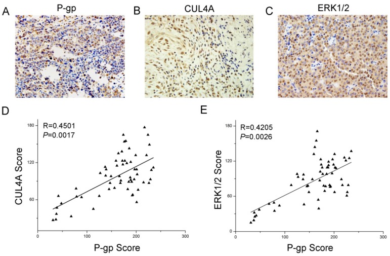
Figure 7.
Expression and relationship of P-gp, CUL4A and ERK1/2 in breast cancer patient samples. Immunohistochemical analysis of P-gp (A), CUL4A (B) and ERK1/2 (C) expression in breast cancer tissues (59 cases; magnification of ×400). (D) P-gp expression was positively correlated with CUL4A expression in breast cancer tissues. (E) P-gp expression was positively correlated with ERK1/2 expression in breast cancer tissues.

