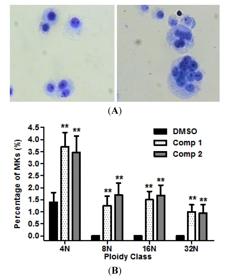
Figure 4.
Compounds induced megakaryopoiesis in HEL cells. (A) Light microscopy shows that the image of megakarycytes (control and compound 1, left and right respectively) stained by Giemsa; (B) The percentage of megakaryocytes was determined on the basis of the number of endomitosis. Results are representative of at least three independent experiments run in triplicate and expressed as the mean ± SD.

