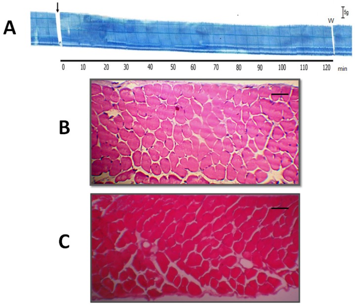
Figure 4.
Histological analysis of muscle damage in PND incubated with or without 7,8,3'-trihydroxy-4'-methoxyisoflavone (TM). Panel (A) shows a representative trace of PND incubated with TM (n = 4). The arrow indicates the addition of TM. Bar = tension of 5 g/cm. W = wash. Panels B and C show cross-sections of diaphragm muscle incubated with Tyrode solution alone (negative control; n = 6) (B) or TM alone (n = 4) (C). Note the normal appearance of the fibers (polygonal aspect and peripheral nuclei) in both panels. Bar = 50 μm in B and C.

