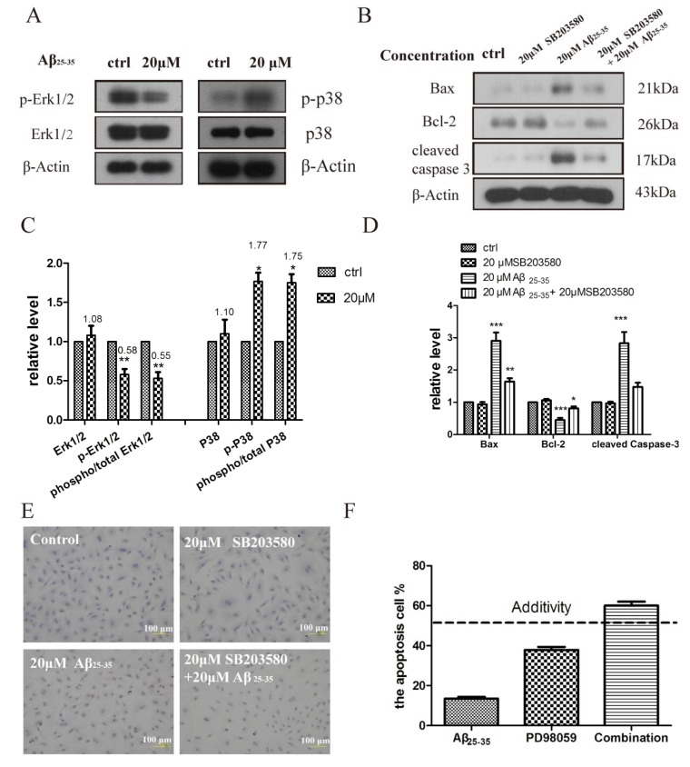
Figure 5.
(A) Aβ25–35 regulates MAPK in isolated rat cardiomyocytes. Immunoblots showing phospho- and total p38, and ERK MAPKs in isolated cardiomyocytes incubated with vehicle, Aβ25–35 (20 μM) for 24 h; (B) Aβ25–35 induced apoptosis protein was inhibited in cardiomyocytes by SB203580 (20 μM) that was added 30 min before the addition of vehicle and Aβ25–35; (C–D) Relative protein levels were quantified by densitometry and shown in the histogram; (E) Aβ25–35 and PD98059 synergistically induced the cardiac myocyte apoptosis. The cardiac myocyte were treated for 24 h with 10 μM Aβ25–35 and 20 μM PD98059. The apoptosis rates were determined by flow cytometry analyses; (F) Aβ25–35 induced TUNEL-positive apoptotic cells was inhibited in cardiomyocytes by SB203580 (20 μM). TUNEL assay was performed, as described in material and methods section, cardiomyocytes cells incubated during 24 h in the absence or in the presence of Aβ25–35 and SB203580. Values are expressed as mean ± SEM of three independent experiments, each in triplicate. * p < 0.05, ** p < 0.01, *** p < 0.001 vs. the control group.

