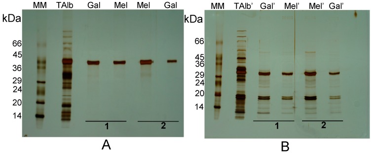
Figure 2.
Electrophoretic profile of the galactose-eluted 42 kDa subunit bound to erythrocyte membranes, performed under non-reducing (A, NR-SDS-PAGE) and reducing conditions (B, R-SDS-PAGE). Two separate but identical sets of erythrocyte membranes were incubated with lupine total albumins (lanes TAlb and TAlb’) and submitted to sequential elution by 0.4 M galactose (elution 1, lanes Gal and Gal’) followed by 0.4 M melezitose (elution 1, lanes Mel and Mel’), or melezitose (elution 2, lanes Mel and Mel’) followed by galactose (elution 2, lanes Gal and Gal’). Molecular masses of standards (MM) are indicated in kDa.

