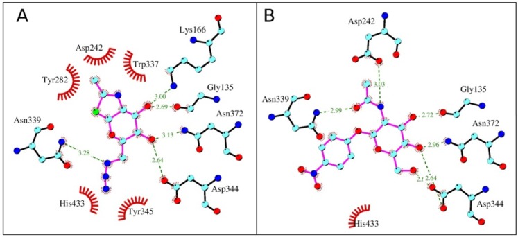
Figure 1.
C-6-Azido-NAG-thiazoline (2) (A) and substrate pNP-β-GlcNAc (B) in the active site of bacterial β-N-acetylglucosaminidase (B. thetaiotaomicron). Color scheme: carbon atom–cyan, oxygen–red, nitrogen–blue, sulfur–green, hydrogens are omitted; hydrogen bonds–green dashed line with marked donor-acceptor length in Ångstroms; residues participating in π-cation interaction are shown schematically.

