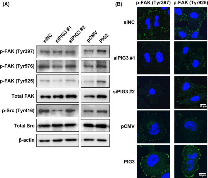
Figure 5.

Fak/Src signaling is stimulated by PIG3. A, A549 cells were transfected with PIG3‐special siRNA (siPIG3) #1, #2 or non‐targeting control siRNA (siNC) and H1299 cells were transfected with PIG3 construct (PIG3) or empty vector (pCMV). Then the total cell lysates were subjected to western blot to analyze the phosphorylation of FAK and Src as described in the Materials and Methods. B, Transfected A549 and H1299 cells were fixed and subjected to immunofluorescence staining using p‐FAK (Tyr397) and p‐FAK (Tyr925) primary antibodies, followed by Alexa‐488 conjugated secondary antibodies (green), coupled with DAPI staining (for nucleus, blue). Scale bar = 10 μm
