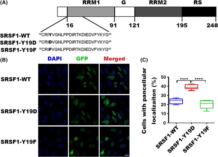
Figure 2.

Localization of WT and mutant serine/arginine‐rich splicing factor 1 (SRSF1). A, Modular structure of SRSF1 and mutants in RNA recognition motif 1 (RRM1). The tyrosine 19 residue (Y19, shown in in boldface type) is phosphorylated, and we mutated it to aspartic acid (D) or phenylalanine (F). B, Indirect immunofluorescence of HeLa cells transfected WT or mutant GFP‐tagged SRSF1. Panels in the right column show the merged images of GFP and DAPI signals. C, Statistics for cells with pancellular (both nucleus and cytoplasm) localization of SRSF1. Three independent experiments were carried out and 3 views were chosen randomly in each experiment. Bar represents SEM. Total number of GFP‐positive cells is 5000 for GFP‐tagged WT or mutant SRSF1. ****P < .0001
