
Figure 1.
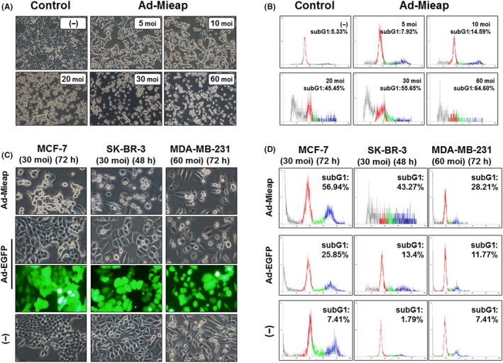
Mieap‐induced cell death in breast cancer cells occurs in a multiplicity of infection (moi)‐dependent manner. A, After 72 h of Ad‐Mieap infection, morphological changes were captured (100× magnification). Cell nuclei were fragmented, and the cell number was decreased in a moi‐dependent manner. B, Adenovirus‐infected cells (MCF‐7) were collected and subjected to FACS analysis. Moi number and percentage of the subG1 fraction (apoptotic cells) are shown in the upper right of each diagram. C, Morphological changes in 3 breast cancer cell lines (MCF‐7, SK‐BR‐3 and MDA‐MB‐231) 48‐72 h after infection at a moi of 30‐60 with Ad‐Mieap. Ad‐Mieap‐infected cells were decreased in number and size. D, After incubation for the indicated time, cells were evaluated by flow cytometry. The subG1 fraction was elevated after Ad‐Mieap infection. The subG1 fractions were 56.94% for MCF‐7 (30 moi, 72 h) and 43.27% for SK‐BR‐3 (30 moi, 48 h). In contrast, most MDA‐MB‐231 cells remained attached, with a subG1 fraction of 28.21%