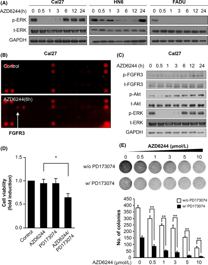
Figure 1.

MEK inhibitor induced an ERK‐activity rebound and fibroblast growth factor receptor 3 (FGFR3) activation. A, Phosphorylated ERK and total ERK protein expression are shown in a representative western blot. Head and neck squamous cell carcinoma (HNSCC) cell lines were treated with 0.5 μmol/L AZD6244 or 0.1% DMSO as a vehicle control for different durations. AZD6244 was replaced with fresh media at the indicated times. GAPDH was detected as a loading control. B, Phospho‐RTK assay in Cal27 cells treated with AZD6244 for 6 h. Cal27 cells incubated with 0.1% DMSO for 6 h served as a control. C, Representative western blot analysis of FGFR3, Akt, and ERK expression in Cal27 cells after treatment with 0.5 μmol/L AZD6244 for different time periods. Media containing AZD6244 was replaced with fresh media (lacking AZD6244) at the indicated times. GAPDH was detected as a loading control. D, Cell growth was measured in Cal27 cells treated with AZD6244 or PD173074 as an FGFR inhibitor in cell‐viability assays. Cells were treated for 48 h with 0.1% DMSO, 0.5 μmol/L AZD6244 alone, 1 μmol/L PD173074, or 0.5 μmol/L AZD6244 with 1 μmol/L PD173074. Bars represent means ± SEM between replicates (n = 3). Significant differences compared to the corresponding controls, *P < .05. E, Clone‐formation ability of Cal27 cells treated with AZD6244 was evaluated in clonogenic assays. Cal27 cells were treated with a dose gradient of AZD6244 in the absence or presence of 1 μmol/L PD173074 for 14 d and were studied in clonogenic assays. Bars represent means ± SEM between replicates (n = 3). Significant differences compared to the corresponding controls, **P < .01
