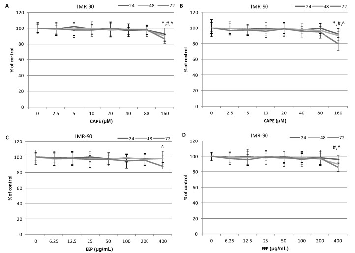
Figure 3.
Cytotoxic effects of EEP and CAPE on the normal lung fibroblast IMR-90 cell line. Cells were incubated with 6.25–200.0 μg∙mL−1 EEP or with 2.5–80.0 μM CAPE for 24, 48 and 72 h. The values represent the mean ± SD of three independent experiments performed in quadruplicate (n = 12). (A) Cytotoxic activity of CAPE against IMR-90 cells. The percentage of cell death was measured using the MTT cytotoxicity assay. (B) Cytotoxic activity of CAPE against IMR-90 cells. The percentage of cell death was measured using the lactate dehydrogenase (LDH) cytotoxicity assay. (C) Cytotoxic activity of EEP against IMR-90 cells. The percentage of cell death was measured using the MTT cytotoxicity assay. (D) Cytotoxic activity of EEP against IMR-90 cells. The percentage of cell death was measured using the LDH cytotoxicity assay. Results are presented as the means of cytotoxicity ± SD. *, ^, # indicate statistically-significant differences compared to the control: * after 24 h of incubation, # after 48 h and ^ after 72 h.

